
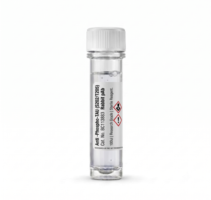
Anti-Phospho-TAU (S202/T205) Rabbit pAb 100ul
Advance your neurobiology research with our high-affinity Anti-Phospho-TAU (S202/T205) Rabbit polyclonal antibody. Designed for superior specificity and sensitivity, this 100ul pack size reagent ensures consistent, reproducible results in your western blot and immunohistochemistry experiments. Unlock deeper insights into tauopathy mechanisms and neurodegenerative disease studies with this reliable, high-performance research tool today.
Product successfully added to the Quote List

Description
Introduction
The Anti-Phospho-TAU (S202/T205) Rabbit pAb is a premium-grade research antibody meticulously developed to detect phosphorylated tau protein sites S202 and T205. Essential for neuroscientific investigations, this antibody provides the precision needed to identify pathological changes associated with Alzheimer’s disease and other related tauopathies in biological samples.
Key Features
– Optimized for high specificity at phosphorylation sites S202 and T205
– Supplied in a convenient 100ul pack size for laboratory efficiency
– Rabbit polyclonal format ensuring robust signal amplification
– Validated for consistent performance in standard immunoassays
– High purity formulation to minimize background noise during imaging
– Stable storage profile to maintain long-term antibody integrity
Benefits
This antibody serves as a critical diagnostic and analytical marker for researchers studying protein aggregation and neurodegeneration. By accurately targeting the S202/T205 phosphorylation sites, it allows for clear visualization of tau pathology. Using this reagent reduces experimental variability, saves valuable time during protocol optimization, and provides the reliable data quality required for peer-reviewed publications and clinical research advancements.
Usage / How to Use
This product is intended for laboratory research use only. Typically, the antibody is diluted in a blocking buffer according to your specific protocol—commonly used for Western Blotting, Immunohistochemistry (IHC), or Immunofluorescence (IF). After membrane blocking or tissue preparation, the primary antibody is incubated with the sample to allow for target binding, followed by appropriate washing and secondary antibody detection steps.
Why Choose This Product
– Unmatched sensitivity for detection of low-abundance phosphorylated targets
– Rigorously tested to ensure batch-to-batch consistency for reproducible data
– Developed specifically for the rigorous demands of modern neurobiology labs
– Exceptional value per microliter, supporting sustainable high-volume research
– Dedicated technical support and clear documentation for easy integration into existing workflows
Additional information
| brands | |
|---|---|
| capacity |
General Inquiries
There are no inquiries yet.



Reviews
There are no reviews yet.